KIS-1 人恶性B细胞淋巴瘤细胞株 BioVector® KIS-1 Human Malignant B-Cell Lymphoma Cell Line
- 价 格:¥99860
- 货 号:BioVector® KIS-1
- 产 地:北京
- BioVector NTCC典型培养物保藏中心
- 联系人:Dr.Xu, Biovector NTCC Inc.
电话:400-800-2947 工作微信:1843439339 (QQ同号)
邮件:Biovector@163.com
手机:18901268599
地址:北京
- 已注册
BioVector® KIS-1 人恶性B细胞淋巴瘤细胞株
BioVector® KIS-1 Human Malignant B-Cell Lymphoma Cell Line
第一部分 中文说明
一 产品基本信息与遗传学背景
细胞名称:BioVector® KIS-1
保藏机构货号:DSMZ ACC 947
物种来源:人类 (Homo sapiens)
性别与年龄:女性,54岁(Japanese 种群背景)
组织与疾病背景:该细胞株建立于1991年,源自一名患有恶性非霍奇金B细胞淋巴瘤(Malignant non-Hodgkin B-cell lymphoma)的54岁日本女性患者的胸腹水(Pleural effusion)组织。临床病理学最初将其归类为具有独特分化表型的间变性大细胞淋巴瘤或免疫母细胞样突变株。
核心致癌分子与转录异常特征:
独特的染色体异位重排:该细胞株在遗传学上表现为复杂的核型特征,其中最具有标志性的是携带染色体易位 t(1;14)(q21;q32)。这一重排将 1 号染色体上的特定基因片段强行置于 14 号染色体免疫球蛋白重链(IGH)增强子的绝对驱动之下。
BCL9 与原癌基因异常激活:通过 t(1;14)(q21;q32) 易位,直接导致位于 1q21 位点上的 BCL9 基因(B-cell lymphoma 9) 发生严重的转录失控和病理性高表达。高丰度的 BCL9 蛋白作为 Wnt/beta-catenin 信号通路的核心辅助转录因子,过度激活下游的原癌靶基因网络,直接促进了淋巴瘤细胞的恶性无限增殖和凋亡抗性。
免疫表型谱(Immunophenotype):
阳性表面标记:CD19、CD20、CD22、CD38、CD45、HLA-DR、sIgM、sIg-kappa。
阴性表面标记:CD3、CD5、CD10、CD23、CD30。
生物安全级别:1级(BSL-1)。经多重 PCR 检测,EBV、HBV、HCV、HIV-1/2、HTLV-1/2 均为阴性。
二 细胞形态学与培养环境
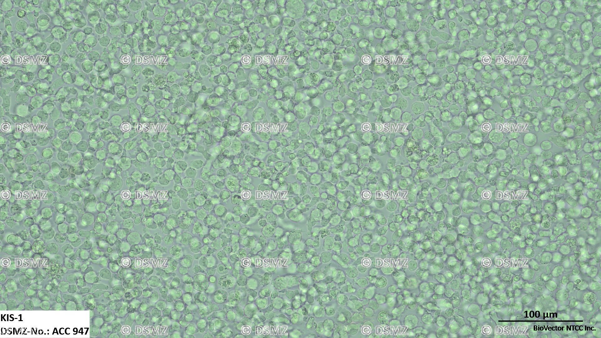
形态学特征:展现典型的恶性B细胞淋巴瘤或大细胞淋巴瘤形态特征。在低倍显微镜下,细胞大小相对不均一,主要以单个散在的圆形细胞或数十个细胞松散黏附形成的悬浮团块(Single cells and loose aggregates)形式在培养基中生长。
生长模式:完全悬浮生长。
群体倍增时间(Doubling Time):大约 36 至 48 小时。
标准完全培养基配方:
基础培养基:80% 至 90% BioVector® RPMI-1640 培养基。
维持添加:10% 至 20% 优质热灭活胎牛血清(h.i. FBS)。注:对于生长脆弱、易形成自发性碎片的悬浮淋巴突变株,早期复苏强烈建议采用 20% 的血清浓度加压维持。
1% Penicillin-Streptomycin 双抗溶液。
物理培养参数:37摄氏度恒温、5% 二氧化碳、空气饱和湿度。
三 细胞传代与复苏标准操作步骤
初始接种与常规传代操作(Subculturing Protocol):
起始接种密度:当首次从液氮罐复苏或大比例稀释该细胞时,初始细胞密度必须维持在较高的起点,推荐设定在每毫升约 $0.4 \times 10^6 \sim 0.6 \times 10^6$ 个活细胞。强烈建议先在 24 孔板或 T25 培养瓶中进行局部高密度垂直维持,切勿过度稀释。
日常维持密度窗口:在日常扩增循环中,应将细胞密度严格控制在 每毫升 $0.5 \times 10^6$ 至 $1.5 \times 10^6$ 个活细胞 之间。
传代执行:当悬液中的活细胞密度接近每毫升 $1.5 \times 10^6$ 个的饱和峰值或培养基因乳酸堆积明显变黄时需要进行分裂传代。由于是完全悬浮生长,直接通过低速离心(150 g,5分钟)收集细胞,去除旧基后加入新鲜预热的完全培养基重悬。常规传代比例为 1比2 至 1比3,通常每 2 到 3 天常规补液或传代一次。
冻存细胞复苏:
快速将冻存管自液氮罐中取出,移入 37 摄氏度 BioVector® 水浴锅中持续高频摇晃解冻,在 1 到 2 分钟内令其极速融化。
将解冻的细胞液缓慢移入含有 5 毫升 预热高血清完全培养基的无菌离心管中,以 150 g 离心 5 分钟。
彻底吸除含有 DMSO 冻存液的上清,使用新鲜的完全培养基轻轻重悬细胞沉淀,并按照上述推荐的高密度启动参数接种培养,置于箱内静置,前 24 小时避免频繁晃动。
四 核心科研应用方向
t(1;14) 易位与 BCL9/Wnt 通路在淋巴瘤发病中的分子机制研究:BioVector® KIS-1 是国际上研究携带经典 t(1;14)(q21;q32) 染色体易位的恶性 B 细胞淋巴瘤极其罕见且极其珍贵的内源性标准工具株。主要用于阐明 BCL9 异常激活如何协同 beta-catenin 信号在 B 细胞淋巴瘤发生、恶性演进及骨髓侵袭过程中的转录重塑机制。
新型 Wnt/beta-catenin/BCL9 阻断剂的小分子药效学筛选:由于该细胞株天然高表达 BCL9 蛋白且依赖此通路维持增殖,它是评估和筛选新型 BCL9-beta-catenin 蛋白质相互作用(PPI)小分子抑制剂、Wnt 信号通路特异性靶向药或抑制剂的理想体外药效动力学评价模型。
针对 B 细胞系表面靶点的现代免疫疗法(CAR-T/双特异性抗体)效能评估:该细胞表面稳定且高丰度地表达成熟 B 细胞标记物 CD19、CD20、CD22 以及 HLA-DR。常被广泛用作肿瘤靶细胞,在体外效效比测定和细胞毒性释放实验中,用于量化评估和筛选新一代抗 CD19/CD20/CD22 双特异性单抗、抗体偶联药物(ADCs)以及 CAR-T、CAR-NK 细胞的靶向溶瘤能力。
PART 2 ENGLISH SECTION
I General Information and Genetic Background
Cell Line Name: BioVector® KIS-1
Repository Catalog Number: DSMZ ACC 947
Species Origin: Human (Homo sapiens)
Sex and Age of Donor: Female, 54 years old (Japanese population profile)
Tissue and Disease Background: Established in 1991 from the malignant pleural effusion fluid of a 54-year-old Japanese female patient diagnosed with Malignant Non-Hodgkin B-cell Lymphoma. Clinical and pathological screening originally designated this line within the large-cell anaplastic or immunoblastic B-lymphoma categories exhibiting distinct lineage markers.
Core Oncogenic Markers & Transcriptional Aberrations:
Landmark Chromosomal Translocation: Characteristically accommodates the pathognomonic reciprocal chromosomal translocation t(1;14)(q21;q32). This rearrangement places genetic loci from chromosome 1 under the direct downstream influence of the strong Immunoglobulin Heavy Chain (IGH) enhancer complex on chromosome 14.
BCL9 Overexpression & Wnt Pathway Activation: The t(1;14) event specifically results in the heavy transcriptional deregulation and marked pathognomonic overexpression of the BCL9 (B-cell lymphoma 9) gene located at the 1q21 locus. Elevated BCL9 acts as an essential co-activator of the Wnt/beta-catenin transcriptional framework, heavily accelerating oncogenic target genes that govern unrestricted B-cell expansion and anti-apoptotic defense circuits.
Immunophenotypic Profile:
Positive Cell Surface Antigens: CD19, CD20, CD22, CD38, CD45, HLA-DR, sIgM, and sIg-kappa.
Negative Cell Surface Antigens: CD3, CD5, CD10, CD23, and CD30.
Biosafety Level: BSL-1. Validated negative via multiplex PCR tracking for EBV, HBV, HCV, HIV-1/2, HTLV-1/2, and MLV viral genomes.
II Morphological Attributes and Cultivation Media
Morphology: Displays distinctive architecture typical of malignant high-grade B-lymphoblastic variants. Under standard phase-contrast microscopy, cells present as pleomorphic spherical units growing either as isolated individual suspension cells or as multi-cellular loose suspension aggregates.
Growth Mode: Strict suspension growth.
Population Doubling Time: Approximately 36 to 48 hours.
Standard Complete Growth Medium Formulation:
Basal Medium: 80% to 90% BioVector® RPMI-1640 medium.
Routine Supplements: 10% to 20% premium BioVector® Heat-Inactivated Fetal Bovine Serum (h.i. FBS). Note: Elevating serum concentrations to 20% is highly recommended during immediate post-thaw recovery phases to rescue fragile suspension clusters.
1% Penicillin-Streptomycin solution.
Physical Incubation Parameters: Regulated strictly at 37 degrees Celsius under a humidified atmospheric layer containing 5% Carbon Dioxide.
III Subculturing and Thawing Protocols
Initial Seeding Setup and Routine Passaging Schedule:
Seeding Initialization: When initializing a fresh culture from a cryovial or initiating post-split expansion, always seed the cell suspension at a high density baseline, preferably between $0.4 \times 10^6$ and $0.6 \times 10^6$ viable cells/mL. It is highly recommended to confine the culture volume in a 24-well plate or small flask to optimize paracrine conditioning signaling loops.
Core Maintenance Density Window: During routine long-term maintenance cycles, keep the active expanding cell density restricted tightly between $0.5 \times 10^6$ and $1.5 \times 10^6$ cells/mL.
Subculturing Routine: Split the culture when the biomass parameters reach or approach the saturation ceiling of $1.5 \times 10^6$ cells/mL. Because it expands purely in suspension, enzymatic trypsinization is completely unnecessary. Pellet the suspension by spinning at 150 g for 5 minutes, decant the spent broth, and redistribute the cell mass in fresh complete medium at recommended split ratios of 1:2 to 1:3 every 2 to 3 days.
Cryovial Thawing and Recovery:
Retrieve the cryovial from cryogenic storage parameters and plunge it into a 37 degrees Celsius BioVector® water bath with continuous rapid manual agitation, achieving complete liquefaction within 1 to 2 minutes.
Transfer the cell slurry slowly into a sterile centrifuge tube carrying 5 mL of pre-warmed complete growth medium and spin at 150 g for 5 minutes.
Decant the supernatant completely to clean out toxic residual DMSO traces, and gently resuspend the cell pellet in fresh, serum-enriched complete growth medium, establishing the recommended high density seeding configuration.
IV Strategic Research Applications
Unraveling t(1;14) Translocation and BCL9/Wnt Transcriptional Axis Oncogenesis: BioVector® KIS-1 serves as an invaluable, rare endogenous reference model naturally harboring the reciprocal t(1;14)(q21;q32) chromosomal translocation. It is widely used to dissect how aberrant BCL9 expression interfaces with the beta-catenin transcriptional complex to drive Wnt pathway hyperactivation and dictate aggressive non-Hodgkin lymphoma progression.
Screening Targeted Small-Molecule Wnt / BCL9 Inhibitors: Given its dependence on the functional BCL9-beta-catenin axis for structural survival, this cell line functions as an optimized pharmacological tool to screen small-molecule inhibitors of the BCL9-beta-catenin protein-protein interaction (PPI), map novel target degraders, and evaluate therapeutic synergy parameters.
Validating Anti-B-Cell Modern Immunotherapies (CAR-T, CAR-NK, or Bi-specific Formats): Because KIS-1 cells stably present major mature B-lineage surface determinants including CD19, CD20, CD22, and HLA-DR on their outer membranes, they function as validated target lines in standard cytotoxicity assays. They are routinely deployed to evaluate the binding affinity, target engagement, and oncolytic efficiencies of multi-specific monoclonal antibodies, Antibody-Drug Conjugates (ADCs), and engineered CAR-T or CAR-NK cells.
BioVector NTCC质粒载体菌株细胞蛋白抗体基因保藏中心
电话:400-800-2947
工作QQ/微信同号:1843439339
网址http://www.biovector.net
- 公告/新闻